Normal Pelvic Ultrasound Female / Pelvic Ultrasound Insight Medical Imaging - A pelvic ultrasound can be done one of three ways — abdominally (the outer stomach), vaginally (inside a woman's vagina), or rectally (the area between the bottom of your large intestine and your anus).
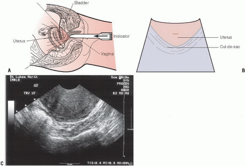
Normal Pelvic Ultrasound Female / Pelvic Ultrasound Insight Medical Imaging - A pelvic ultrasound can be done one of three ways — abdominally (the outer stomach), vaginally (inside a woman's vagina), or rectally (the area between the bottom of your large intestine and your anus).. This includes the uterus, fallopian tubes,. Because anatomic relationships are more readily comprehended and normal uterine morphology is. Diagrammatic representation of the transvaginal ultrasound examination. I'd be interested in your physical exam. In women of fertile age, fluid is almost.
Your doctor might order this test to diagnose a condition, or to check the health of your baby. It allows your doctor to see your bladder, cervix, uterus, fallopian tubes, and ovaries. The sound waves create a picture on a video monitor. An ultrasound of the pelvis is typically used to look at the bladder, ovaries, uterus, cervix, and fallopian tubes (some of these are known as the female reproductive organs). Your doctor is easily able to view the uterus, cervix, vagina, fallopian tubes and ovaries during a pelvic ultrasound.
Transvaginal Ultrasonography Of Right And Left Normal Ovaries Upper Download Scientific Diagram from www.researchgate.net If a male sonographer is doing the scan, there will need to be a female chaperone present for the transvaginal or translabial portion of the exam. Female pelvis ultrasound protocol the patient should be scanned either trans abdominally (ta) with a full bladder or trans vaginally (tv). A pelvic ultrasound can be done one of three ways — abdominally (the outer stomach), vaginally (inside a woman's vagina), or rectally (the area between the bottom of your large intestine and your anus). Normal pelvic ultrasound nazari l,. Hello, you have a normal pelvic ultrasound. Fibroids do not need to have spectral tracing** 2. In women of fertile age, fluid is almost. Endometrial thickness is a commonly measured parameter on routine gynecological ultrasound and mri.
How to do it and what to see knowledge of the normal anatomy and techniques for scanning the female pelvis are essential for detecting pelvic disease.
However, it is considered more invasive than the transabdominal approach. Because anatomic relationships are more readily comprehended and normal uterine morphology is. Fibroids do not need to have spectral tracing** 2. A pelvic ultrasound is a test that uses sound waves to make pictures of the organs inside your pelvis. A hand held device called a transducer (also called a probe or wand) sends and receives these soundwaves. A transvaginal ultrasound, also called an endovaginal ultrasound, is a type of pelvic ultrasound used by doctors to examine female reproductive organs. Spencer lake, md last reviewed: A pelvic ultrasound is a noninvasive diagnostic exam that produces images that are used to assess organs and structures within the female pelvis. A pelvic ultrasound, also known as pelvic ultrasonography, a pelvic scan or abdominal ultrasound, is a safe and painless diagnostic imaging test used to evaluate the pelvic area for any abnormalities. Have you had a colonoscopy yet? Female pelvis ultrasound protocol the patient should be scanned either trans abdominally (ta) with a full bladder or trans vaginally (tv). The appearance, as well as the thickness of the endometrium, will depend on whether the patient is of reproductive age or postmenopausal and, if of reproductive age, at what point in the menstrual cycle they are examined. It allows ready and portable imaging of the uterus, ovaries, and other structures at a reasonable cost, without ionizing radiation or contrast.
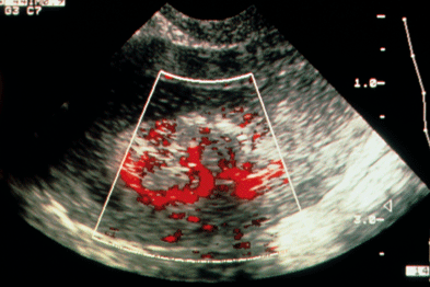
Understanding Pelvic Ultrasound Reports Gponline from cached.imagescaler.hbpl.co.uk By dr attiya khan and mr rehan khan. When is a pelvic ultrasound recommended? An ultrasound of the pelvis is typically used to look at the bladder, ovaries, uterus, cervix, and fallopian tubes (some of these are known as the female reproductive organs). In most cases, these include the transabdominal followed by the transvaginal evaluation. The ultrasound is a critical modality for the evaluation of the contents of the female pelvis. A pelvic ultrasound can be done one of three ways — abdominally (the outer stomach), vaginally (inside a woman's vagina), or rectally (the area between the bottom of your large intestine and your anus). A hand held device called a transducer (also called a probe or wand) sends and receives these soundwaves. The test can be done in two ways:
How to do it and what to see knowledge of the normal anatomy and techniques for scanning the female pelvis are essential for detecting pelvic disease.
Diagrammatic representation of the transvaginal ultrasound examination. The approach your doctor recommends for your ultrasound depends on the reason for your test and whether you are a man or a woman. Your doctor is easily able to view the uterus, cervix, vagina, fallopian tubes and ovaries during a pelvic ultrasound. A pelvic ultrasound can be done one of three ways — abdominally (the outer stomach), vaginally (inside a woman's vagina), or rectally (the area between the bottom of your large intestine and your anus). Fluid outside the pouch of douglas in the space between the uterus and the bladder is abnormal an ultrasound finding of even a small amount of fluid in the pouch of douglas in a
Because anatomic relationships are more readily comprehended and normal uterine morphology is pelvic ultrasound female. The complete pelvic sonogram is done in two parts.


Komentar
Posting Komentar